Frequently asked questions
Is it too late to have cartilage reconstruction done?
No and yes. It is of course a clinical decision made only after thorough consideration. Most of the traumatic lesions can be treated in working age patients with pretty good results. In top level elite athletes it is more a question of missed season due to long rehab and protective period before returning to professional sports. And yes, definitely there are cases where only total join replacement is clinically relevant surgical approach.
Does it interfere if the very same area has already been treated with other cartilage repair technique?
This depends on techniques planned. Microfractures or cell cultures are not used if there is a previous microfracture or drilling on the very same area. If deep osteochondral reconstruction is planned, then earlier operations do not prevent reconstruction. There are several deep techniques, examples are included on this site.
How do I know if my symptoms are related to cartilage problem?
Swelling, exercise related pain and stiffness are classical signs of cartilage problem, but symptoms can also include sharp pain and locking in more acute trauma. For a long time, cartilage problems have been considered only after all other possible reasons have first been ruled out. With current imaging methods cartilage issues can be seen and diagnosed earlier, just like meniscus problems.
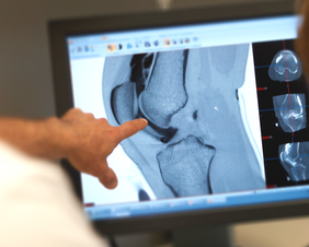
I already have an MRI done, with no signs of cartilage problem. Can I still a have a cartilage lesion?
Accuracy of MRI in detecting cartilage lesions is highly variable depending on machinery and imaging center, review articles suggest that MRI can detect 70-80% of cartilage lesions. In practice, every fourth patient is perhaps missed in routine knee MRI. This problem is mostly related to limited imaging time and spatial resolution in magnetic resonance imaging technique.
Can I get more accurate imaging done?
MRI can be optimised for cartilage imaging in many ways, and there are MRI imaging centers specialised in cartilage diagnostics. If even more accurate imaging is needed, then cone beam CT (CBCT) scanning is nowadays available. It can produce exceptionally high resolution of 0,2 mm in any given direction. Imaging is absolutely fast, taking only 18 seconds.
Can I follow my cartilage condition with MRI?
MRI is a highly sensitive imaging method in cartilage diagnostics. Cartilage layers are thin convex and concave 3D volumes, which are difficult to reconstruct from slice based MRI imaging data. Results may vary depending on patient positioning, imaging device and many other things.
CBCT is an X-ray based imaging method. How high risk is radiation dose in CBCT?
CBCT technology has originally been developed for safe imaging of head and neck area containing some very sensitive organs. Imaging of peripheral joint is even smaller risk. Radiation dose in typical joint imaging is comparable to 3 to 5 native plain X-rays, far less than in large scale CT-scanners. CBCT imaging requires injection of contrast media in joint, so iodine allergy is a contraindication.
Why is my joint swelling after exercises?
Exact mechanisms are not known in every detail. In general, joint is lubricated by joint fluid produced by synovium in your joints. If anything irritates joint, more liquid is pumped in to lubricate joint. Cartilage lesions are one reason for this kind of irritation leading to swelling, worsening after exercise, and relieving after rest.
Why is it difficult to go up or down stairs?
Patellofemoral joint is a common area for cartilage lesions. Especially knee cap side lesions interfere with going up stairs or squatting. Trochlea, or sulcus, is part of the thigh bone and facing knee cap. Cartilage lesion on this side are more often symptomatic when coming down the stairs.
Is it true that some of the cartilage lesions can be reconstructed using small arthroscopic portals?
Yes, actually we perform all our surgery arthroscopically, except patella reconstruction. Patella lesions require a short (5 to 7 cm) arthrotomy on either medial or lateral side. Ankle (TC-joint) is treated totally arthroscopically, it is very rare that larger incisions are needed on that area.
What if I need osteotomy or ligament surgery?
Especially in knee area it is important to rule out predisposing factors leading to cartilage issues. If clinical visit indicates, mechanical axis imaging of the whole lower extremities is indicated. It is important to consider treatment of these alignment and ligament problems first, otherwise reconstructed cartilage is not in safe.
If there is no answer to your specific question here, please do not hesitate to ask us using our contact form or sending an email to info@cartilagehelp.com
|
|
The content of this website is provided for information only and is not intended to be used for diagnosis or treatment or as a substitute for consultation with your own doctor or a specialist.Email addresses supplied are provided for basic enquiries and should not be used for urgent or emergency requests, treatment of any knee injuries or conditions or to transmit confidential or medical information.
|